Hydrodynamic cavitation is mostly an undesired phenomenon in turbomachinery since it limits the performance of the fluidic system, causes catastrophic damage, flow choking and acoustic noise, and lowers the efficiency. However, it could be used it for good means in small scale.
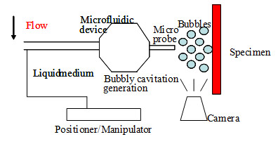
In this study, continuous bubbly cavitating flows with the design of a microfluidic device (bubble generator) were generated. The emerging bubbles were exposed to a small target area (kidney stones, cells in culture, human tissues). Macroscopic, microscopic and nanoscopic (e.g. molecular) changes in the target area using both biology and surface characterization methods were monitored.
This size of micro-orifice was chosen to enable visualization of microbubbles emerging due to bubbly cavitating flow by a simple digital camera. The material involves a sheet of protection and has high resistance to corrosion thus it can be used in applications that require sensitivity.
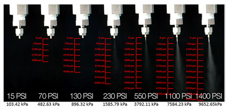
The tests were conducted by applying different inlet pressure values at the inlet. The flow rate was controlled with a fine-metering valve. Various inlet pressure values were applied during the tests in order to observe the result at increasing pressure differences until bubbly cavitating flow pattern is obtained.
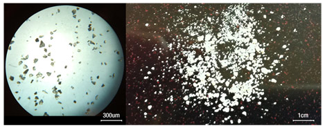
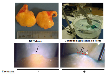
It was observed in kidney stone experiments that the targeted application of the erosion with micro scale hydrodynamic cavitation causes the fracture of the kidney stones within a short time of 30 min. Moreover, micro scale bubbly cavitation caused cell death in two different cancer cell types in culture, and cell death kinetics depended on the exposure time to cavitation. In addition to cancerous cells, the destructive effect of hydrodynamic cavitation was investigated on human tissues. Hydrodynamic cavitation exposure resulted in a deep cavity on the targeted area of BPH tissue without giving any damage to the surrounding area. All these new findings support the idea of usage of hydrodynamic cavitation as a novel therapeutic method for ablating tumor tissues.

References:
Üzüşen, D., Perk, O.Y., Demir, E., Oral, Ö., Ekici, S., Ünel, M., Gözüaçık, D., and Koşar, A., "Assessment of probe-to-specimen distance effect in kidney stone treatment with hydrodynamic cavitation," ASME Journal of Medical Devices, Under Review.
Itah, Z., Kosar, A., Sesen, M., Ekici, I.D., Ekici, S., and Gozuacik, D., "Hydrodynamic Cavitation Kills Prostate Cells and Ablates Benign Prostatic Hyperplasia Tissues," Experimental Biology and Medicine, 238 (11), 2013.
Perk, O.Y., Şeşen, M., Gözüaçık, D., and Koşar, A., "Kidney Stone Erosion by Hydrodynamic Cavitation and Consequent Kidney Stone Treatment," Annals of Biomedical Engineering, 40, pp. 1895-1902, 2012.
Koşar, A., Sesen, M., Oral, O., Itah, Z. and Gozuacik, D., "Bubbly Cavitating Flow Generation and Investigation of its Erosional Nature for Biomedical Applications", IEEE Transactions on Biomedical Engineering, 58, pp. 1337-1346, 2011.